by Markus W. Risinger and Ryan T. McGuire
Investigators commonly point to retinal hemorrhages, or retinal bleeding, to distinguish child abuse from accidental injury or underlying medical conditions. Courts rely on expert testimony identifying retinal hemorrhages as evidence of child abuse so frequently that the diagnosis has almost become shorthand for an abuse finding. But are retinal hemorrhages such clear and reliable indicators of abuse that the inquiry can simply stop there? Are all retinal hemorrhages the same? Although everyone would like to have a single medical finding that can differentiate between traumatic/non-traumatic or accidental/non-accidental injuries in children, the case for retinal hemorrhages as a forensic “lodestar” is not so certain.
What are retinal hemorrhages?
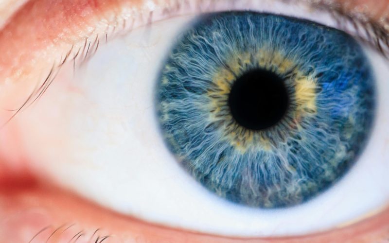
To understand the significance of retinal hemorrhages in child abuse investigations, one must first understand the diagnosis itself. In the simplest terms, retinal hemorrhage describes bleeding in part of the eye. The retina is a layer of tissue, located in the back of your eye, made up of specialized cells that convert light into electrical signals to transmit to the brain. The brain interprets those signals to form the image you see when you observe the world around you. The retina is essential for vision but very sensitive. It can be damaged by excessive light exposure (like staring into the sun), high pressure within the eye or pressing against the retina from inside the skull, and traumatic injury. Like other tissues within the body, the retina bleeds when damaged, resulting in visible spots and discoloration when viewed from outside the eye. Doctors can use several devices, such as a retinoscope, to view, measure, and photograph the retina and identify retinal hemorrhages.
Are all retinal hemorrhages the same? How are they categorized?
The retina is not just a single layer of cells. Rather, it contains numerous layers of cells that serve different purposes. Some parts of the retina are directly exposed to light entering the eye while others connect to other structures, like the optic nerve, to convert light to useful information and transmit it to the brain. “Retinal hemorrhage” is a broad term used to describe any bleeding in the retina or even in adjacent tissues, but it is not very specific as to which tissues are bleeding or in which area of the eye. The retina is not a flat object; it is three-dimensional and surrounds most of the internal structures of the eye.
The Kellogg Eye Center classifies retinal hemorrhages based on their anatomical location and appearance. Dot and blot hemorrhages appear as small, round spots when tiny blood vessels deep within the retina burst. Flame hemorrhages earn their name from their flame-like appearance on examination and occur when blood vessels near the retina’s surface rupture within the nerve fiber layer. Boat-shaped or pre-retinal hemorrhages develop when larger surface blood vessels rupture and blood pools between the retina and the eye’s clear vitreous gel, creating a distinctive curved appearance. Subretinal hemorrhages form beneath the retina when blood vessels in the underlying choroid layer rupture, typically near the macula. Finally, vitreous hemorrhages happen when blood vessels on the retina’s surface or abnormal tissue growths bleed into the eye’s clear vitreous gel, significantly blurring vision. [1]
What can cause retinal hemorrhages?
Trauma, disease, and various underlying medical conditions and genetic disorders can cause or worsen retinal hemorrhages. Understanding the full spectrum of potential causes is crucial for proper diagnosis and legal assessment.
Trauma
Child abuse can cause retinal hemorrhages through significant trauma that impacts the retinal areas. Accidental trauma can also cause retinal hemorrhages. Falling on a hard surface—even from just a distance of a couple of feet—can cause retinal hemorrhages, but it is nearly impossible to know what caused the fall from studying the injury to the eye alone. Intentional infliction of child abuse is not a necessary condition for retinal hemorrhages, nor can the source of trauma be easily identified by the mere existence of hemorrhages, the type of hemorrhages, or the severity of hemorrhages without reference to other physical findings. Birth trauma is also a recognized cause of retinal hemorrhages observable for a period after a child is born. Birth is a significant traumatic event, but no one could call a birth child abuse.
Disease
Various diseases can produce retinal hemorrhages. As noted by David Rossiaky for healthline.com, examples include “diabetic retinopathy, hypertensive retinopathy, retinal vein occlusion, Anemia, leukemia, bacterial endocarditis, sickle cell anemia, preeclampsia, high altitude retinopathy (caused by exposure to extreme high altitudes), [and] lupus.” [2] Some of these causes can be studied and ruled out in specific cases, but the full extent of hemorrhages caused, prolonged, or worsened by disease in children is unknown.
Medical Conditions and Genetic Disorders
Coagulopathy is a condition where the blood’s ability to clot is impaired. This can be caused by genetic disorders like hemophilia and Von Willebrand disease. [3] Coagulopathy can also result from acute sources, like certain medications. Regardless of how it comes about, a clotting disorder can contribute significantly to retinal findings.
Retinal Hemorrhages in Very Young Children
Retinal hemorrhages are particularly significant indicators when found in very young children because many age- and lifestyle-related conditions that commonly cause retinal hemorrhages can be ruled out. Young children, especially non-mobile infants, will not have experienced some of the known causes of retinal bleeding in adults (e.g., preeclampsia). Additionally, traumatic events in very young children should theoretically be easier to identify and document, particularly for infants who cannot yet move independently. This process of elimination makes retinal hemorrhages in young children a more focused diagnostic indicator, though not an exclusive one.
What is the legal value of retinal hemorrhage?
It is tempting to look for a medical diagnosis or physical observation that is exclusive to abuse because that would make the investigative process universally easier. However, scientists have only scratched the surface of understanding the retina and its ailments. Further research must be done, and scrutiny is warranted whenever a medical expert claims a high degree of certainty about what caused a child’s condition. The lack of certainty is why the law requires evidence to satisfy a burden of proof. Retinal hemorrhages, even severe ones, are not perfectly diagnostic of a particular mode of injury despite how readily an investigator or factfinder may want to accept that kind of explanation. Yet medical authorities and courts still consider retinal hemorrhages as an important indicator of child abuse when found in conjunction with other evidence.
Sources:
[1] Retinal Hemorrhages : Ophthalmoscopic Abnormalities : The Eyes Have It
[2] Retinal Bleeding: Symptoms, Causes, Diagnosis, and Treatment
[3] Management of bleeding and coagulopathy following major trauma: an updated European guideline – PMC
Markus Risinger is an attorney at Woodnick Law and has been practicing family law since graduating from ASU’s Sandra Day O’Connor College of Law. He practices and teaches administrative law, juvenile law, family law, criminal law, and appeals.
Ryan T. McGuire is a law clerk at Woodnick Law PLLC and a second-year student at the Sandra Day O’Connor College of Law. He leverages his master’s degree in philosophy to produce thorough legal research and writing, with a focus on family law matters. Ryan brings a strong foundation in legal analysis and academic interests spanning family, criminal, and business law.


